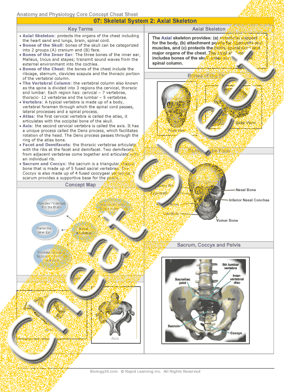
The axial skeleton provides: (a) structural support for the body, (b) attachment points for ligaments and muscles, and (c) protects the brain, spinal cord and major organs of the chest. The axial skeleton includes bones of the skull, inner ear, chest and spinal column.
Bones of the Skull: Can be categorized into two groups: (A) Neurocranium and (B) Splanchnocranium. With the exception of the mandible, all the bones of the skull are joined together by sutures.
The Neurocranium includes the following bones:
- Frontal bone: makes up the forehead and part of the eye orbits and part of the nasal cavities.
- Parietal bones: here are 2 parietal bones, which articulate together and form the roof of the cranium.
- Temporal bones: one on either side of the skull, contain the inner ear. These bones also provide a foramen (canal) for the major blood supply to the brain, the carotid artery and jugular vein.
- Occipital bone: makes up the back and floor of the cranium. The brainstem passes through this bone and then continues as the spinal cord.
- Ethmoid bone: forms the front part of the cranial floor, part of the eye orbits, and contains the ethmoid sinuses.
- Sphenoid bone: contains the sphenoidal sinus cavity. Has a unique depression called the sella turcica, which houses the pituitary gland.
- Palatine Bone: these bones are at the back of the roof of the mouth. They form the wall of the nasal cavities and the floor of the eye orbit.
Bones of the Face: The bones of the face (Splanchnocranium) are 14 in total. They are: 2 nasal, 2 maxilla, 2 zygomatic, 2 lacrimal, mandible, 2 palatine, 2 inferior nasal conchae and vomer.
- Lacrimal Bone: is the smallest bone of the face, from part of the inside wall of the eye orbit.
Nasal Bone: the two nasal bones meet in the middle and this forms the bridge of the nose.
Inferior Nasal Conchae: these bones form the lateral wall of the nasal cavity and cause the inhaled air to swirl and be filtered.
- Vomer Bone: is a triangular shaped bone that forms part of the nasal septum.
Zygomatic Bone: it is a paired bone, which makes up the lower eye orbit and is frequently referred to as the cheekbone.
- Maxilla Bone: the largest bones of the face; they form together to make the whole upper jaw. These bones hold the upper teeth.
- Mandible Bone: the strongest bone of the face; it forms the lower jaw and holds the lower teeth. It is the only bone of the skull that moves.
- The hyoid bone is a bone in the neck, which does not articulate with any other bone. Muscles of the neck support it and it provides support for the root of the tongue; it is involved in the production of speech.
Bones of the Inner Ear: The bones of the inner ear are called the (a) Malleus (hammer), (b) Incus (anvil) and (c) Stapes (stirrup). These bones function together to transmit sound waves from the external environment to the fluid filled cochlea.
- Malleus (hammer): The malleus, or hammer, is a hammershaped bone that is attached to the incus. It is attached to the inner surface of the eardrum and, therefore, it moves as the eardrum vibrates in response to incoming sound.
- Incus (anvil): is an anvil-shaped bone in between the malleus and the stapes. It is the bridge that connects the incoming sound waves to the inner ear.
- Stapes (stirrup): The stapes, or stirrup, transmits the sound vibrations from the Incus to the oval window. The oval window connects the inner ear bones with the cochlea.
Bones of the Chest
Clavicles (or collar bones) are long bones, which support the ribcage and shoulder joints. The clavicles provide an attachment for the scapula and rotate when the arm is moved forward. There are twelve ribs in the rib cage, 10 pairs that are joined to the sternum and spine and 2 floating pairs. The ribs protect the underlying organs and assist in respiration.
- Scapula: also known as the shoulder blade; it is a pair of broad flat bones that connect the arm bone with the clavicle.
- Sternum: also known as the breastbone; it is a long flat bone in the center of the chest. It connects to the ribs via cartilage and completes the rib cage. It has three portions, from the top downward: (A) Manubrium, (B) Body and (C) Xiphoid Process.
The Vertebral Column:
- Cervical: The cervical region is the first portion of the spinal column and is made up of 7 vertebras. The first and second vertebrae are unique, and they are called the atlas and the axis.
- Thoracic: The thoracic region of the vertebral column is located in the chest. It contains 12 vertebrae and is connected to the lumbar region of the spine.
- Lumbar: The lumbar region of the vertebral column is the last main portion of the vertebral column and is located in the lower back. It contains 5 vertebrae and is connected to the pelvis, through the sacrum and coccyx.
- Sacrum: The sacrum is a triangular shaped bone that is made up of 5 fused sacral vertebrae. It articulates with and provides a strong foundation for the pelvis.
- Coccyx: The coccyx is also a triangular shaped bone that is made of 4 fused coccygeal vertebrae, and is also known as the tailbone. It is attached to the sacrum by cartilage, and this allows some movement between them and shock absorbance.